
Revision Eyelid Surgery in Seoul, Korea
Customized design for the most important part of your facial impression, restoring your natural beauty.
Revision Eye Surgery
Revision surgery is not a single standardised procedure. A crease that is too high or too low, a loosened line, significant scarring, bilateral asymmetry — the underlying cause and the appropriate solution differ entirely from case to case. Because tissue architecture is altered by previous surgery and the internal state cannot be fully predicted in advance, it is often necessary to directly visualise and restore damaged structures intraoperatively. Ultimately, the outcome of revision surgery comes down to the surgeon's experience and judgement.
Quick Facts
| Procedure | Revision Eye Surgery · Revision Eye Surgery |
|---|---|
| Surgeon | Dr. Seungsoo Kim (Board-certified Plastic Surgeon, 10+ yrs) |
| Time | 1–2 hours |
| Anesthesia | IV Sedation |
| Hospitalization | Not required |
| Stitch Removal | 5–7 days post-op |
| Follow-up Visits | 3 or more visits |
| Recovery | 2 weeks or more |
| Best For | Crease placed too high, creating a heavy or unnatural appearance ('sausage lids') · Crease placed too low, making it barely visible · Crease loosening with a faded or indistinct line · Noticeable asymmetry between the two eyes · Thickened or prominent scarring from a previous procedure · Unsatisfactory results despite multiple previous surgeries |

Check Point Highly Recommended For
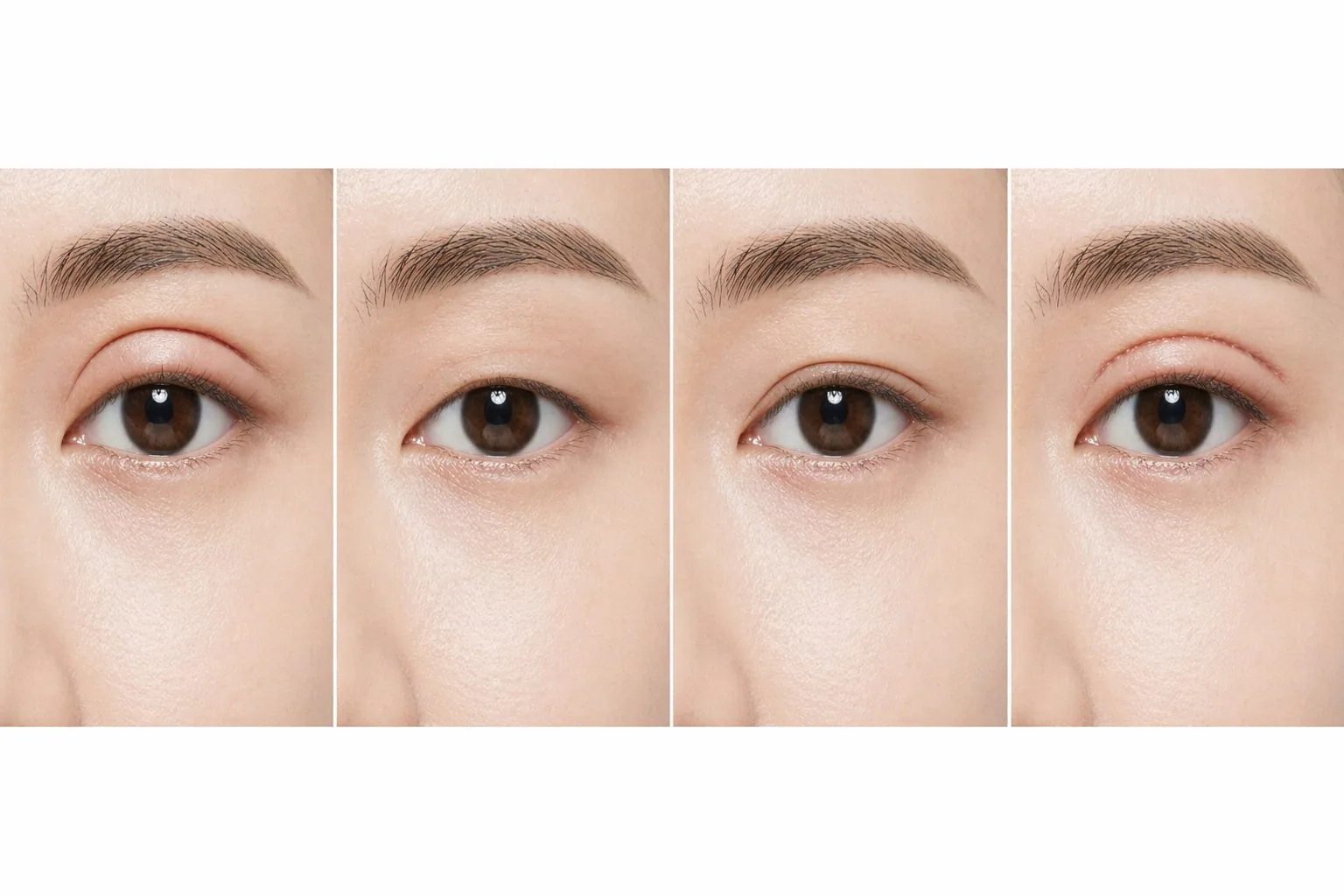
Revision Eye Surgery Procedure
Precise Diagnosis
Previous surgical history and root causes of unsatisfactory results are identified, and the five key factors — amount of residual skin, fat volume, muscle tone, bilateral difference, and existing crease height — are measured to classify the case type.
Tailored Surgical Plan
Based on the case type (crease too high, loosening, scarring, or asymmetry), dissection extent, restoration approach, and need for combined procedures (skin excision, fat grafting, ptosis correction) are determined.
Adhesion and Scar Dissection with Structural Restoration
Adhesions and scar tissue altered by previous surgery are meticulously addressed, and any damaged structures — levator palpebrae superioris, orbicularis oculi, etc. — are restored under direct visualisation.
Crease Reformation and Fixation
A new crease height and shape suited to the patient's current anatomy are designed and precisely secured. Bilateral asymmetry is addressed with fine individual adjustment on each side.
Tension-Free Closure
Meticulous tension-free suturing prevents additional scarring. Revision patients are followed up with scheduled visits to monitor the healing progress.
4 Representative Cases for Revision Eye Surgery — Different Causes Require Different Solutions
The term 'revision surgery' covers a wide spectrum. The dissection extent, restoration approach, and expected recovery differ entirely depending on the problem being corrected — which is why precise diagnosis is always the first priority.
Crease Placed Too High ('Sausage Lids')
The crease height was set too aggressively in the previous procedure, or the skin and muscle were over-tightened. The existing adhesion line is released through an incision and a new, lower crease is formed. If the upper skin has also descended and folded over, a brow lift may be considered in conjunction.
Crease Loosening / Too Low
Point adhesion from a burial suture procedure has loosened, or the crease was originally set too low to be clearly visible. The incision method is used to create direct adhesion, minimising the risk of recurrence, and the crease is redesigned to suit the current eyelid thickness and skin condition.
Significant Scarring or Crease Distortion
Aggressive tarsal and muscle anchoring or imprecise suturing in the previous procedure has resulted in thick scar tissue. Scar tissue and adhesions are carefully dissected and the crease is reformed using tension-free suturing. This is the case type that most significantly increases the complexity of revision surgery.
Bilateral Asymmetry
Differences in crease height, shape, or degree of lid opening between the two sides. A discrepancy in levator function between the two eyes is often present, requiring incision-based ptosis correction with fine individual adjustment on each side.
5 Factors Evaluated During Revision Surgery Assessment
In revision surgery, 'what the patient's eyes look like now' matters more than 'what procedure was previously performed.' Even among cases of the same type — such as crease loosening — the surgical plan changes depending on the combination of these five factors.
Eye Surgery Procedures — At a Glance
The right method depends on lid thickness, sagging, eye proportion, and revision needs.
| Procedure | Area | Time | Anesthesia | Recovery | Key Technique |
|---|---|---|---|---|---|
| Natural Adhesion Double Eyelid Natural Adhesion | Upper eyelid (double-fold line) | 30 minutes | IV Sedation / Local | From day 3–4 | Micro-tunnel + natural adhesion |
| Incision Double Eyelid Incision Method | Upper eyelid (skin/muscle/fat) | 1 hour | IV Sedation / Local | From 1 week post-op | Incision + tissue trim + line fixation |
| Canthoplasty Canthoplasty | Medial / lateral / lower canthus | 30 minutes | Local / IV Sedation | From 1 week post-op | CPF anchoring + skin redraping |
| Ptosis Correction Ptosis Correction | Upper eyelid (levator muscle) | 1 hour | IV Sedation | From 1 week post-op | Levator strengthening |
| Under-Eye Fat Repositioning Under Eye Fat Repositioning | Lower eyelid (conjunctiva/fat) | 30 minutes – 1 hour | IV Sedation | From 1 week post-op | Conjunctival fat repositioning |
| Revision Eye Surgery Revision Eye Surgery | Eye revision | 1–2 hours | IV Sedation | 2 weeks or more | Adhesion release + redesign |
Click a row to open that procedure.
The AVA Difference
Natural Adhesion Technique
Tissue is repositioned along natural planes and secured at multiple points.
Designed to reduce loosening and align with the patient's intrinsic contour.
Levator Aponeurosis Ptosis Correction
Ptosis is addressed by shortening and refixing the levator aponeurosis,
restoring natural eyelid flow.
Patient Satisfaction Defines Completion
Surgery is complete not when sutures are removed,
but when the patient smiles in front of the mirror.
Revision eyelid surgery corrects problems left by a previous double eyelid operation, such as crease height, loosening, scarring or asymmetry, with a different approach for each cause.
Who it’s not for
We advise caution within six months of the previous surgery, while swelling and adhesions are still settling; when skin and tissue have already been removed extensively, leaving little room to revise; and when the expectation is the same as for a first operation.
What we suggest instead
We diagnose the cause first, whether height, loosening or asymmetry, and in many cases a partial correction is enough.
Pick a surgery date to see stitch removal, return to daily life and the earliest flight home as calendar dates. Confirmed at consultation.
- Arrive in Seoul · consultationD-1Fri, Sep 25
- SurgeryD+0Sat, Sep 26
- Stitch removalD+7—
- Back to daily lifeD+14Wed, Sep 30
- Earliest flight homeD+14Thu, Oct 1
- Major swelling settledD+90Mon, Oct 26
Individual recovery and combined procedures change the dates.
Cost depends on the extent of surgery, anesthesia and any combined procedures, and is quoted at consultation. The same criteria apply to every patient.
See current offersQ&A
Post-op Instructions
- Do not rub the surgical area strongly for about a week.
- Avoid alcohol and smoking for at least 2 weeks as they delay recovery.
- Swelling and bruising vary individually but will gradually improve.
- Light walking or stretching helps reduce swelling.
Curious About This Procedure?
Our head surgeon provides a one-on-one assessment and a tailored treatment plan just for you. Results and recovery may vary by individual.