
Alar Reduction in Seoul, Korea
Not just raising the nose, but meticulously designing the natural angle from forehead to the tip.
Alar Reduction
The alar base accounts for approximately one-third of the frontal impression of the nose. Beyond simply reducing width, the more fundamental concern is scarring — the alar region is subject to constant movement from the facial muscles, meaning incision placement and suture tension are what ultimately determine the outcome. AVA categorises alar morphology into four types (lateral bulk · medial nostril size · alar base width · alar ptosis) and designs the incision position differently for each, minimising the risk of visible scarring.

Check Point Highly Recommended For
Alar Reduction Procedure
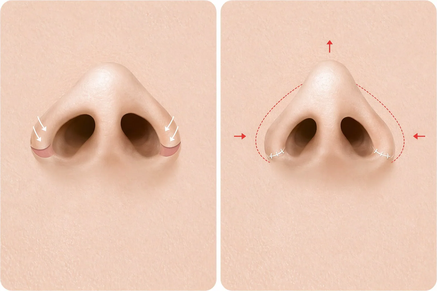
CASE 01. Thick Lateral Alar Wall (Lateral Incision)
Suited for cases where the lateral alar wall itself is thick and wide. A portion of tissue is excised along the inner border of the alar-cheek junction and sutured to reduce the alar width from the front. Because the incision follows the natural contour of the groove, it becomes almost imperceptible over time.
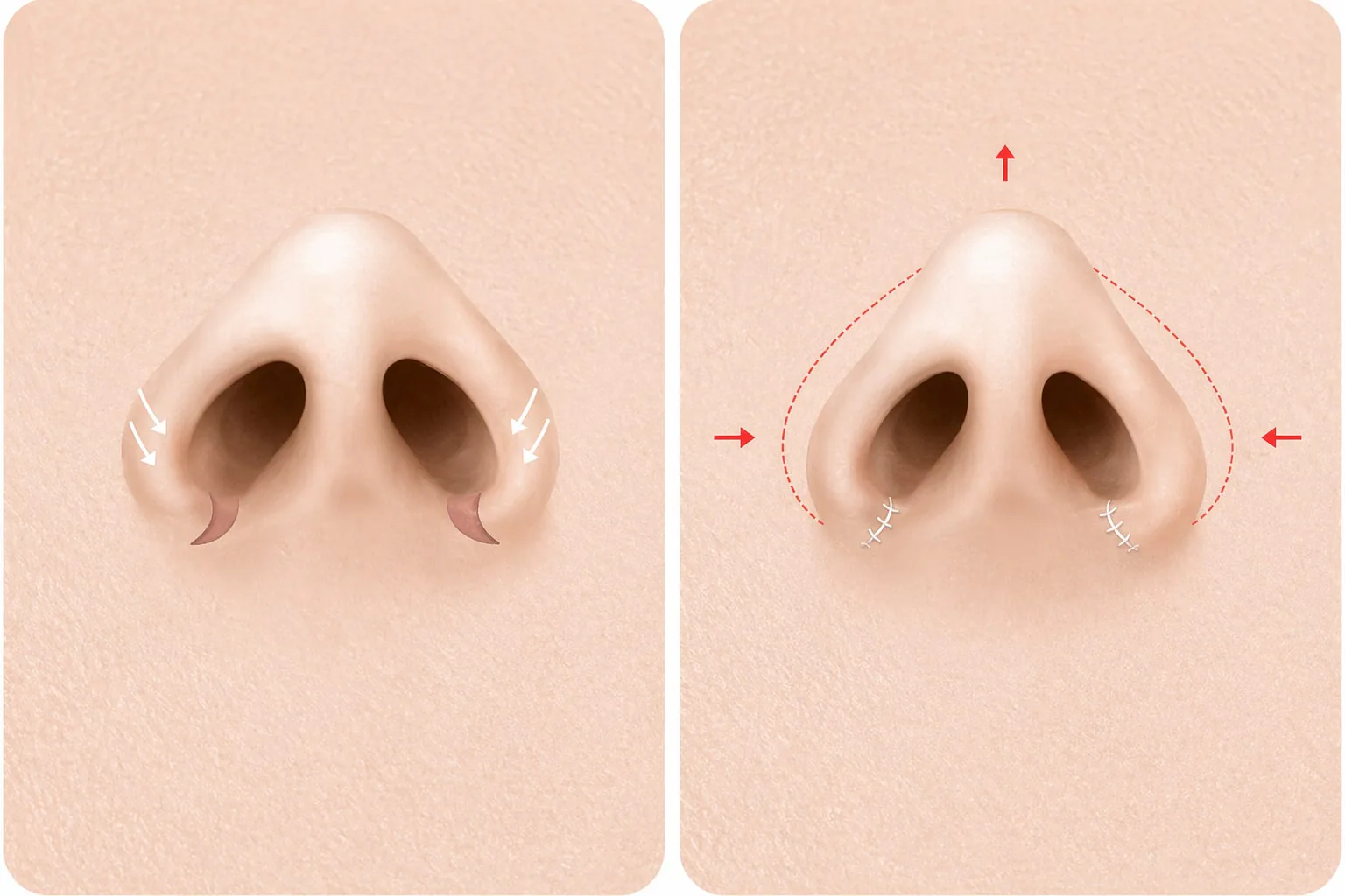
CASE 02. Large Nostrils (Medial Incision)
Suited for cases where the lateral alar wall is relatively thin but the nostril opening itself is large. Tissue is excised from within the nostril sill to reduce nostril size, leaving no external scar — making this the lowest-risk option in terms of visible scarring.
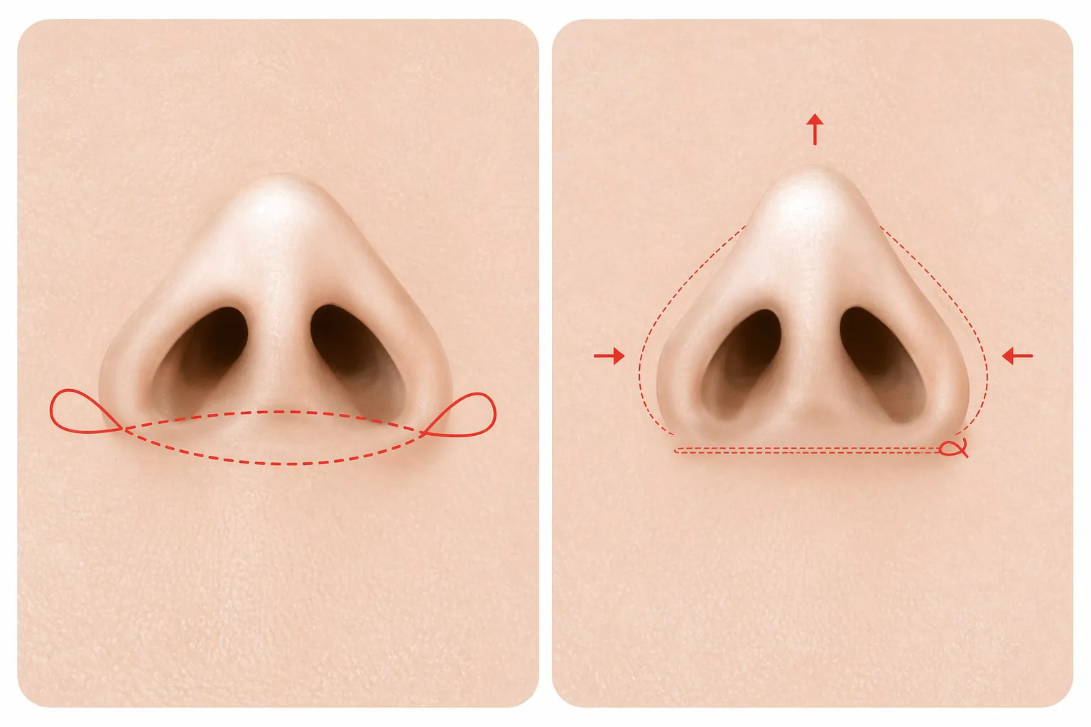
CASE 03. Wide Alar Base (Cinching Suture, No Incision)
Suited for cases where lateral or medial excision is not required but the overall alar base width needs to be narrowed. A permanent non-absorbable suture is passed through the inner alar tissue on each side and cinched together to draw the alar bases towards the midline. No external incision is made, so there is no external scar.
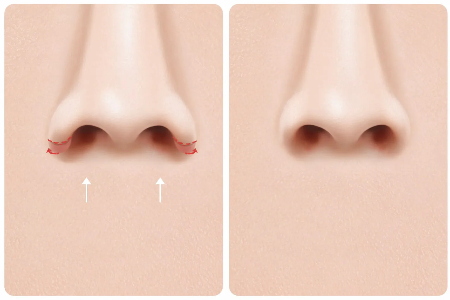
CASE 04. Alar Ptosis (Alar Lift)
Suited for cases where the alar base width is acceptable but the alar rim droops downward — making the nostrils appear elongated, particularly when smiling. Tissue at the superior alar is partially excised and sutured to lift the drooping rim. When indicated, this is combined with septal extension to achieve integrated correction of the tip-to-alar relationship.
The AVA Difference
Autologous Rib Cartilage Rhinoplasty (Especially Revision)
Reduces implant-related concerns by reconstructing nasal architecture
using the patient's own 6th–7th rib cartilage.
Park-Kim-Lee Revision Algorithm
A Stranc classification-based revision algorithm (Archives of Craniofacial Surgery, 2017)
that considers structural defects and respiratory function together.
Patient Satisfaction Defines Completion
Surgery is complete not when sutures are removed,
but when the patient smiles in front of the mirror.
Alar reduction reduces the size or width of wide nostrils, with incision placement and suture tension designed for the nostril shape.
Who it’s not for
Not advised as a stand-alone operation when the nose looks wide because the tip is low rather than because the nostrils are wide, since narrowing them alone then looks unnatural.
What we suggest instead
Raising the tip often makes the nostrils look narrower on their own, so we consider it together with rhinoplasty.
Pick a surgery date to see stitch removal, return to daily life and the earliest flight home as calendar dates. Confirmed at consultation.
- Arrive in Seoul · consultationD-1Fri, Sep 25
- SurgeryD+0Sat, Sep 26
- Stitch removalD+7Sat, Oct 3
- Back to daily lifeD+7Sat, Oct 10
- Earliest flight homeD+7Sat, Oct 10
- Major swelling settledD+90Mon, Oct 26
Individual recovery and combined procedures change the dates.
Cost depends on the extent of surgery, anesthesia and any combined procedures, and is quoted at consultation. The same criteria apply to every patient.
See current offersQ&A
Post-op Instructions
- Do not rub the surgical area strongly for about a week.
- Avoid alcohol and smoking for at least 2 weeks as they delay recovery.
- Swelling and bruising vary individually but will gradually improve.
- Light walking or stretching helps reduce swelling.
Curious About This Procedure?
Our head surgeon provides a one-on-one assessment and a tailored treatment plan just for you. Results and recovery may vary by individual.