Epicanthoplasty, 45° Upper & Lateral/Lower Canthoplasty Anatomy — Periosteal Fixation & Safety Limits
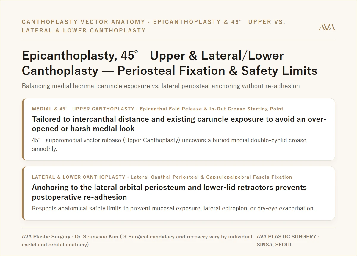
- Epicanthoplasty and 45° upper canthoplasty are not about cutting skin wider; they release the fibrous epicanthal band and preseptal orbicularis tension to balance intercanthal proportion, lacrimal caruncle exposure, and the 45° superomedial starting fold of an in-out or semi-out double eyelid.
- Preventing lateral/lower canthoplasty re-adhesion ("sticking back") or mucosal ectropion requires anchoring the lateral canthal structure to the inner lateral orbital rim periosteum and securing the lateral lower tarsus via the capsulopalpebral fascia (lower-lid retractors) rather than skin-only snipping.
- Strict anatomical safety boundaries must be respected: when the lateral orbital pocket is shallow, lower scleral show is already present, or lower-lid laxity and dry-eye symptoms are significant, excessive lateral/lower extension is avoided to protect eyelid closure and ocular surface health.
"Dr. Kim, I want my eyes to look more open and refreshed, but will medial epicanthoplasty expose too much pink tissue or make my eyes look too close together? And is it true that lateral or lower canthoplasty either closes back up or pulls the lower eyelid outward?" — In canthoplasty, natural results depend not on maximizing millimeter opening, but on matching the medial epicanthal tension vector (horizontal vs. 45° superomedial) and anchoring lateral/lower structures to the orbital periosteum and retractors strictly within safe anatomical limits.
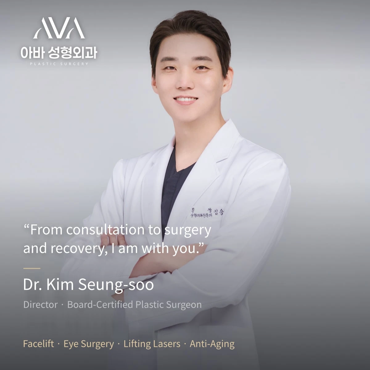
Hello, I am Dr. Seungsoo Kim, Chief Director and Board-Certified Plastic Surgeon at AVA Plastic Surgery in Sinsa, Gangnam, Seoul.
When patients consult for non-incisional double eyelid surgery or incisional blepharoplasty, one of the most common companion questions is whether canthoplasty (medial, 45° upper, lateral, or lower eyelid descent) is appropriate for their eye frame. While double eyelid and ptosis surgery refine vertical eye height, canthoplasty refines horizontal width and medial-lateral inclination. Because over-correction can compromise both expression and ocular comfort, let us examine the two anatomical axes that govern safe canthoplasty.
01. Axis 1 (Medial & 45° Upper Vector): Medial Epicanthoplasty vs. 45° Upper Canthoplasty
The medial epicanthal fold ("Mongolian fold") is not merely excess skin; it is a three-dimensional web formed by tight preseptal orbicularis oculi muscle fibers and dense fibrous bands pulling inferomedially across the inner corner of the eye.
- Horizontal Medial Epicanthoplasty (Medial Vector):
- Indicated when intercanthal distance is wide relative to facial width and a heavy epicanthal fold covers the lacrimal caruncle (the small pink structure at the inner corner), making the eyes appear short or constricted.
- Preventing Over-Correction: If a patient already has a narrow intercanthal distance or prominent caruncle exposure, aggressive horizontal opening creates a harsh, cross-eyed appearance and visible step-off scars. We release only the internal fibrous tension band and redrape the skin without tension, preserving balanced caruncle visibility.
- 45° Upper Canthoplasty (Superomedial Vector):
- Indicated when the distance between the eyes is already balanced, yet an oblique fold pulls downward over the upper-inner corner—burying the starting point of an in-out or semi-out double eyelid crease and giving the medial eye a hooked contour.
- By releasing tension along the 45° superomedial vector rather than pulling horizontally toward the nasal bridge, we open the upper-medial curve smoothly without narrowing the intercanthal gap.
02. Axis 2 (Lateral & Lower Vector): Why Lateral & Lower Canthoplasty Require Periosteal & Retractor Fixation
Patients who experienced "re-adhesion" after a previous lateral canthoplasty elsewhere almost always underwent a superficial skin-and-conjunctiva snip. Because the lateral canthus is under constant medial elastic tension every time you blink, unanchored incisions naturally zip back together during wound healing.
- Lateral Canthoplasty with Lateral Orbital Periosteal Fixation: We first palpate the depth of the lateral orbital rim pocket. When anatomical space permits, we release the lateral retinacular tension and anchor the new lateral canthal angle directly to the internal periosteum of the lateral orbital rim—preventing postoperative re-adhesion while maintaining sharp lateral canthal apposition against the globe.
- Lower Canthoplasty ("Glamorous Line" / Lower Eyelid Descent) via Capsulopalpebral Fascia: For patients with an upwardly slanted lateral lower lid, approaching through the transconjunctival plane and anchoring the lateral third of the lower tarsal border to the capsulopalpebral fascia (lower eyelid retractors) gently lowers the lateral curve without external lower-lid skin scarring.
03. Anatomical Safety Boundaries: Preventing Ectropion, Mucosa Exposure & Dry Eye
As a board-certified plastic surgeon, my foremost rule in canthoplasty is knowing when not to operate or when to limit the extent of opening:
- Contraindications & Conservative Limits: When the lateral orbital rim lies flush against the globe with no recessed pocket, when lower scleral show ("three-white eyes") or lower-lid laxity is already present, or when severe dry-eye symptoms exist, aggressive lateral or lower canthoplasty risks lateral ectropion (red mucosal exposure), blunted "U-shaped" lateral angles, eyelash loss, and chronic ocular irritation. In such anatomy, we advise against over-extension.
- Canthal Revision & Restoration: When prior over-resection has left excess caruncle exposure medially or mucosal eversion laterally, revision requires anatomical layer-by-layer reconstruction of the medial orbicularis/canthal band or lateral canthal tendon anchoring rather than simple skin closure.
04. Recovery Timeline, Chemosis Care & Recommended Stay in Seoul
- Days 5 to 7 (Suture Removal): Fine micro-sutures at the medial or lateral canthus are removed between postoperative days 5 and 7.
- Weeks 1 to 2+ (Subconjunctival Edema / Chemosis Resolution): After lateral or lower canthoplasty, temporary clear swelling of the conjunctiva (chemosis) or mild redness is common for 1 to 2 weeks (occasionally longer in revision or combined cases) and resolves with prescribed eye drops and cool compresses. International patients should plan at least 7 to 10 days in Seoul so suture removal and early conjunctival healing can be checked in person before flying home.
- Months 1 to 6 (Scar Maturation): Mild pinkness at the canthal margin fades progressively over 3 to 6 months into the natural mucocutaneous shadow.
※ Clinical Notice: Candidacy and recovery for medial epicanthoplasty, 45° upper canthoplasty, lateral/lower canthoplasty, and canthal revision vary by individual intercanthal distance, lacrimal caruncle exposure, globe prominence, lateral orbital rim depth, and lower-lid tone. Potential risks and side effects include bleeding, infection, swelling, temporary subconjunctival edema (chemosis), dry-eye irritation, hypertrophic scarring, partial re-adhesion, asymmetry, over-exposure of the lacrimal caruncle, or lateral mucosal exposure/ectropion. Always complete a 1:1 in-person examination with a board-certified plastic surgeon before deciding on surgery.
Book Your 1:1 Consultation with Dr. Seungsoo Kim
Whether you are flying in from overseas or living in Korea, Dr. Seungsoo Kim personally oversees every step from initial consultation to surgery and recovery:
- 📅 Book a 1:1 Zoom Video Pre-Consultation or Seoul Clinic Visit (English Booking Hub) →
- 📖 Read Dr. Kim's Mother's Deep-Plane Facelift & Eyelid Surgery 2-Month Recovery Log →
- ✨ View Deep-Plane Facelift Guide & 1-Year Follow-Up Cases →
- 👁️ View Upper Eyelid, Sub-Brow & Forehead Lift Guide →
- 💬 Chat Directly on WhatsApp (+82 10-3705-3423) →
📍 AVA Plastic Surgery · 4F Insung Building, 603 Gangnam-daero, Seocho-gu, Seoul (1-min walk from Sinsa Station Exit 4) · TEL +82 2-545-1882
※ Surgical & Consultation Notice: 1:1 Zoom video consultations provide a preliminary educational assessment; final surgical candidacy and operative plans are confirmed through an in-person clinical examination by Dr. Seungsoo Kim in Seoul. Post-operative complications such as bleeding, infection, swelling, scarring, asymmetry, or sensory changes may occur, and results and recovery vary by individual.
Frequently Asked Clinical Questions (FAQ)
Q. What is the anatomical difference between medial epicanthoplasty and 45° upper canthoplasty?
Medial epicanthoplasty primarily addresses horizontal webbing across the inner corner and adjusts intercanthal distance and lacrimal caruncle visibility. By contrast, 45° upper canthoplasty releases the diagonal superomedial skin-muscle hooding that presses down on the inner crease, allowing an in-out or semi-out double eyelid line to begin smoothly without over-exposing the inner red caruncle when the eyes are already close-set.
Q. Why do some lateral and lower canthoplasties stick back together or expose red conjunctiva?
Every blink generates sphincter-like orbicularis contraction that pulls a skin-only lateral incision closed—or pulls an unanchored lower lid downward into ectropion. Durable, natural shaping requires deep periosteal fixation inside the lateral orbital rim and controlled lower-lid retractor anchoring, strictly bounded by the depth of the patient’s lateral orbital bone pocket and lower-lid snap-back tone.
Q. What is the recovery timeline and recommended stay in Seoul for four-direction canthoplasty?
Fine sutures are removed on Post-Op Day 5 to 7. Temporary subconjunctival edema (chemosis) or mild redness after lateral/lower canthoplasty typically subsides over 1 to 2+ weeks with lubricating and anti-inflammatory eye drops. International patients are advised to plan at least a 7- to 10-day stay in Seoul (longer for revision or combined eyelid surgery) before flying home.
Read the original Naver blog post ·