Bulbous Nasal Tip Plasty & Alar Base Reduction Anatomy — Cartilage Support vs. 3 Alar Reduction Techniques
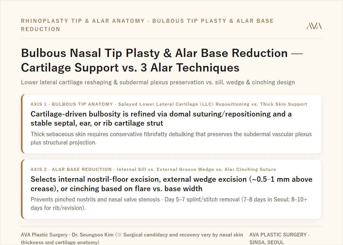
- Correcting a bulbous nasal tip requires distinguishing cartilage-driven bulbosity (wide or weak lower lateral cartilages, LLC) from soft-tissue-driven bulbosity (thick sebaceous skin and subcutaneous fibrofatty tissue), combining domal suturing and structural strut support with conservative debulking that strictly preserves the subdermal vascular plexus.
- Alar base reduction uses the intercanthal-to-alar proportion as a facial harmony reference rather than a rigid numeric target, selecting between internal nostril-sill excision, external alar-groove wedge excision (~0.5–1 mm above the natural crease with deep tension-free sutures), and cinching sutures according to nostril floor width versus lateral alar flare.
- Respecting anatomical safety limits prevents over-thinning necrosis or pollybeak deformity at the tip and avoids "pinched/clothespin" nostrils, step-off scars, or nasal valve stenosis at the alar base.
"Dr. Kim, my nasal tip looks round and heavy in photos and my nostrils flare wide when I smile—will tying the tip cartilage alone make my nose look slimmer, or do I also need alar base reduction? And how do you avoid that artificial 'pinched' nostril look?" — Refining a bulbous tip and wide alar base requires a two-axis anatomical diagnosis: (1) distinguishing cartilage-driven bulbosity from thick sebaceous soft-tissue bulbosity at the nasal tip, and (2) matching the alar reduction technique (internal sill, external wedge, or cinching suture) to nostril floor width vs. lateral flare.
Hello, I am Dr. Seungsoo Kim, Chief Director and Board-Certified Plastic Surgeon at AVA Plastic Surgery in Sinsa, Gangnam, Seoul.
In Asian and revision rhinoplasty consultations, one of the most frequent requests is refining a heavy, rounded ("bulbous") nasal tip alongside wide nostrils. Because the nasal tip lobule and the alar base form an interconnected equilateral triangle when viewed from below, planning structural tip plasty and alar base reduction in isolation can distort lower-third harmony. Let us examine the two anatomical axes that govern safe, natural tip and nostril refinement.
01. Axis 1 (Bulbous Nasal Tip Anatomy): Cartilage-Driven vs. Soft-Tissue-Driven Bulbosity
Two noses that appear similarly round from the outside often have completely different internal anatomy when examined during open structural rhinoplasty:
- Type 1 — Cartilage-Driven Bulbosity (Hypertrophic or Splayed Lower Lateral Cartilages, LLC):
- The overlying skin is thin-to-moderate, but the paired lower lateral cartilages (alar cartilages) are broad, convex, or splayed apart at the dome—creating a wide, boxy, or undefined tip.
- Surgical Principle: Conservative cephalic trim of excess lateral crural width is combined with precise transdomal and interdomal suturing to bring the tip-defining points together, supported on a stable columellar strut or septal extension graft (using septal, ear, or autologous rib cartilage depending on septal strength and revision history).
- Type 2 — Soft-Tissue-Driven Bulbosity (Thick Sebaceous Skin & Subcutaneous Fibrofatty Tissue):
- Even when the underlying cartilage is narrow, a thick sebaceous skin envelope and dense subcutaneous fibrofatty tissue (nasal SMAS layer) mask the underlying definition.
- Preserving the Subdermal Vascular Plexus & Preventing Pollybeak Deformity: Aggressively scraping the underside of the dermis to "thin the skin" risks injuring the subdermal vascular plexus, which can cause ischemic skin necrosis, dimpling, or unpredictable scar contracture. Moreover, if a heavy skin envelope is left without strong structural projection, dead space above the tip fills with scar tissue—creating a supratip fullness known as a pollybeak deformity. Therefore, we perform conservative deep fibrofatty debulking strictly beneath the subdermal plexus and pair it with firm structural cartilage tip projection that tents the thick skin envelope from within.
02. Axis 2 (Alar Base Reduction): Choosing Among Internal Sill, External Groove Wedge & Alar Cinching
While textbook facial canons compare alar base width to the intercanthal distance (the space between the inner corners of the eyes), we treat this proportion as a harmony reference guide rather than a rigid target—accounting for overall facial width, ethnic balance, and the natural narrowing ("tenting effect") that occurs as nasal tip projection increases. Based on the anatomy of the nostril floor and lateral alar lobule, we select from three approaches:
- Internal Nostril Sill Excision (Medial Floor Approach):
- Indications: Best suited when the nostril floor (sill) itself is horizontally wide, with minimal lateral overhanging of the outer alar wall.
- Design Advantage: The excision is confined within the natural nostril sill border, narrowing nostril breadth while keeping the incision concealed inside the nasal vestibule shadow.
- External Alar-Facial Groove Wedge Excision (or Combined Sill + Wedge Reduction):
- Indications: Indicated when thick lateral alar walls curve convexly outward beyond the alar-facial insertion ("alar flare"). When both wide nostril floors and prominent lateral flare exist, a combined sill-and-wedge design balances both dimensions.
- Scar Concealment & Preventing "Pinched" Nostrils: Placing an incision directly inside the deepest point of the sebaceous alar-facial groove can obliterate the natural sulcus and leave a depressed track. Instead, we place the external incision approximately 0.5 to 1 mm superior to the natural alar-facial crease, preserve a subtle curved transition at the sill, and close the deep muscular/dermal layer tension-free before placing fine surface sutures.
- Non-Incisional Alar Cinching Suture:
- Indications & Limits: A cinching suture alone cannot remove thick excess skin or large alar lobules and may relax over time if used as a standalone substitute for tissue excision. It is reserved for mild dynamic alar widening when smiling, or used as an internal muscular adjunct alongside tip projection or sill excision.
03. Anatomical Safety Boundaries: Preventing Clothespin Nostrils & Nasal Valve Stenosis
- Project the Tip Before Resecting the Alar Base: Elevating a low, bulbous tip naturally draws the flaring alar walls inward like pitching a tent pole. Performing aggressive alar wedge resection without adequate tip support flattens the lower lateral curvature, creating an artificial "pinched" or "clothespin" nostril contour.
- Protecting the External Nasal Valve & Airway: Because alar skin excision is irreversible, over-resection can narrow the external nasal valve and cause chronic nasal obstruction (nostril stenosis). We always measure nostril symmetry and airway patency so reduction remains safely within functional limits.
04. Recovery Timeline: Day 5–7 Splint/Stitch Removal & Recommended Stay in Seoul
- Days 5 to 7 (Splint & Suture Removal): External nasal splints and fine columellar/alar sutures are removed in clinic between postoperative days 5 and 7.
- Recommended Stay in Seoul for International Patients: For primary bulbous tip plasty and alar base reduction, we recommend a 7- to 8-day stay in Seoul (and 8 to 10+ days when combined with revision rhinoplasty or autologous rib cartilage reconstruction). Remote photo reviews and 1:1 Zoom consultations provide preliminary educational planning; final surgical plans and exact millimeter measurements are confirmed during your in-person physical examination and palpation in Seoul.
- Weeks 2 to 6+ Months (Tip Definition & Scar Maturation): Major swelling subsides within 10 to 14 days, while thick-skin tip edema softens progressively over 3 to 6+ months (and up to 6 to 12 months in thick-skinned or revision patients).
※ Clinical Notice: Candidacy and recovery for bulbous tip plasty and alar base reduction vary by individual nasal skin thickness, sebaceous gland density, lower lateral cartilage strength, and baseline nostril symmetry. Potential risks and side effects include bleeding, hematoma, infection, prolonged tip swelling, temporary numbness, cartilage visibility or deviation, supratip pollybeak fullness, scar contracture, hypertrophic or depressed alar scars, nostril asymmetry, pinched nostril contour, or external nasal valve narrowing (airway obstruction). Always complete a 1:1 in-person examination with a board-certified plastic surgeon before deciding on surgery.

Book Your 1:1 Consultation with Dr. Seungsoo Kim
Whether you are flying in from overseas or living in Korea, Dr. Seungsoo Kim personally oversees every step from initial consultation to surgery and recovery:
- 📅 Book a 1:1 Zoom Video Pre-Consultation or Seoul Clinic Visit (English Booking Hub) →
- 📖 Read Dr. Kim's Mother's Deep-Plane Facelift & Eyelid Surgery 2-Month Recovery Log →
- ✨ View Deep-Plane Facelift Guide & 1-Year Follow-Up Cases →
- 👁️ View Upper Eyelid, Sub-Brow & Forehead Lift Guide →
- 💬 Chat Directly on WhatsApp (+82 10-3705-3423) →
📍 AVA Plastic Surgery · 4F Insung Building, 603 Gangnam-daero, Seocho-gu, Seoul (1-min walk from Sinsa Station Exit 4) · TEL +82 2-545-1882
※ Surgical & Consultation Notice: 1:1 Zoom video consultations provide a preliminary educational assessment; final surgical candidacy and operative plans are confirmed through an in-person clinical examination by Dr. Seungsoo Kim in Seoul. Post-operative complications such as bleeding, infection, swelling, scarring, asymmetry, or sensory changes may occur, and results and recovery vary by individual.
Frequently Asked Clinical Questions (FAQ)
Q. Why can’t a thick-skinned bulbous nose be fixed simply by scraping away more fat under the tip skin?
Aggressive subdermal scraping risks damaging the subdermal vascular plexus, which can lead to skin necrosis, scar contracture, or a supratip pollybeak fullness when thick skin drapes over a weak frame. Safe correction combines conservative deep fibrofatty trimming (preserving dermal blood supply) with a firm septal, ear, or rib cartilage strut that tents and projects the thick skin envelope.
Q. How do you choose between internal sill excision, external wedge excision, and non-incisional alar cinching?
Internal sill excision narrows a wide nostril floor with the scar hidden inside the sill; external wedge excision (placed ~0.5–1 mm above the alar-facial groove to preserve the natural sulcus) reduces thick lateral alar flare; combined sill-and-wedge reduction addresses both; and non-incisional cinching sutures are reserved for mild dynamic flare or used as an adjunct to tip projection.
Q. When are splints and stitches removed, and how long should international patients stay in Seoul?
The external nasal splint and fine columellar/alar sutures are removed on Post-Op Day 5 to 7. We recommend a 7- to 8-day stay in Seoul for primary tip plasty and alar reduction (and 8 to 10+ days when combined with autologous rib cartilage or revision rhinoplasty), while subtle tip definition continues refining over 3 to 6+ months.
Read the original Naver blog post ·